Selasa, 18 Juli 2006
FIling a mesothelioma lawsuit
The mesothelioma lawyers especially at SimmonsCooper lawsuit have been working with clients diagnosed with mesothelioma and asbestos-related diseases for nearly a decade. In that time their mesothelioma lawyers have represented thousands of individuals from all areas of the United States such as they that work as Missouri Mesothelioma Lawyer. They have seen first-hand the pain a mesothelioma diagnosis can cause and are deeply committed to helping victims and families affected by mesothelioma.
The law firm was built on one principle: to give clients and their families the care and respect they deserve. The mesothelioma lawyers know that while each case is unique, they all have one thing in common: every mesothelioma client is a person whose life has been destroyed by someone else’s negligence.
The SimmonsCooper law firm and team of experienced meshotelioma attorneys are extremely knowledgeable about asbestos and, in particular, mesothelioma. As an experienced nationwide mesothelioma law firm, they have learned that one of the most powerful weapons they can offer clients and prospective clients is information.
They understand that filing an asbestos lawsuit is not a simple process, even under the best of conditions. While no amount of compensation can buy the patients a medical miracle, a mesothelioma attorney can help them afford the mesothelioma treatments they need and eliminate the worry that medical costs are draining their family budget.
Senin, 10 Juli 2006
Considering the right drug rehab facility
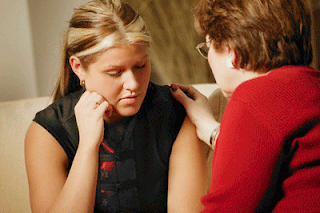
 It’s a decision that can be as daunting as it can be frightening, and it’s most certainly a tough choice for those who have had their lives affected by drugs. Substance abuse rehab centers provide vital support and care for drug addicts in need of treatment, so choosing the right one is crucial to the success and speed of the individual’s recovery.
 Deciding that you or someone you love needs to go to a drug rehab facility is a tough call. Once the decision is made, finding a suitable facility should be as quick and painless as possible, since the 24 to 48 hour period from making that decision, to going out and actually getting help, is the most crucial, if success is to be achieved. Decisions need to be implemented based on solid information.
It is important to find a treatment center that has professional skills and support systems in place that can deal with the addiction. It has to provide an excellent drug treatment program as well as a good drug rehab facility that will build on an individual’s strengths and provide support without judgment or blame. It should guide the individual towards personal accountability, and combined with a detoxification and therapy program, it should enhance a person’s self-confidence to a point where it is possible to integrate back into society.
Type of treatments
There are three common points of all successful drug rehab programs that shoudl always be present : Detoxification, therapy and integration..
The type of treatment that's best for a certain individual will depend largely upon the addiction, the personality type, the addict's degree of dependence, and his social situation. Some centers specialize in specific addictions and others are more general, although one may not be any better than another.
The first thing to decide is whether the individual requires treatment as a resident or as an outpatient. Residential drug treatment is more intense and strict in how the addiction is handled, but it will allow for the best possible care around the clock. Outpatient treatment is typically only successful for a person who doesn’t use daily and still has a job, good family life, etc. In other words, drugs haven’t really ruined his life or relationships yet and he needs a reality check before things get worse. An individual who is actually addicted and not able to control himself or who has experienced consequences as a result of his drug use really has no other option than a residential drug treatment program.
There is, and always has been, a huge debate over which drug treatment programs are more successful. Treatments stem from vastly differing philosophies, and it is here that the conflicts are rooted. What is best for one individual may not be good for another, and only by working with a specialist will a patient be able to determine how best to tackle the addiction.
Withdrawal from drug dependence is something the addict will have to face if he is to overcome his addiction. Some treatments rely on the use of medicinal means to ease withdrawal symptoms, though this method is not right for everyone. Be sure to listen to the advice of the center’s staff; they have been through it before – many of them personally – and they should understand your concerns.
Other types of treatment rely on voluntarily outpatient treatment, which means living and working in a normal way, but with support provided in the form of individual counseling sessions and group classes. Stress is the main factor that triggers a relapse, so learning to cope with this is vital. Twelve Step programs, such as Narcotics Anonymous (N.A.), provide such services.
Finally, there are some types of drug rehab that also attempt to address the underlying causes of addiction, such as family splits, alienation, low employability, mental illness, or living in a community where drug use is prevalent.
This holistic approach to drug rehab provides addicts with new job skills, practical advice, and time to remove themselves from harmful external influences. Such programs often allow patients to stay for a period of time, until they are confident they can function independently without the need to return to drugs.
Kamis, 29 Juni 2006
KERATOSIS SEBOROIK
Keratosis seboroik memiliki banyak manifestasi klinik yang bisa dilihat, dan keratosis seboroik ini terbentuk dari proliferasi sel-sel epidermis kulit. Keratosis seboroik dapat muncul dalam berbagai bentuk lesi, bisa satu lesi ataupun tipe lesi yang banyak atau multipel.
Walaupun tidak ada faktor etiologi khusus yang dapat diketahui, keratosis seboroik lebih sering muncul pada daerah yang terpapar sinar matahari, terutama pada daerah leher dan wajah, juga daerah ekstremitas.
Status dermatologi yang dapat dilihat adalah berbatas tegas, berwarna kecoklatan atau hiperpigmentasi, dan sedikit meninggi disbanding permukaan kulit sehingga penampakan keratosis seboroik seperti tertempel dalam permukaan kulit. Kebanyakan dari keratosis seboroik memiliki permukaan seperti veruka, dengan konsistensi yang halus atau lembut. Walaupun biasanya diameter lesi keratosis seboroik berkisar dalam hitungan beberapa millimeter saja, tetapi ada beberapa lesi yang dapat mencapai ukuran diameter dalam sentimeter. Krusta dan dasar yang inflamasi dapat ditemukan jika lesi terpapar dengan trauma.
EPIDEMIOLOGI
Secara global atau internasional, keratosis seboroik merupakan tumor jinak pada kulit yang paling banyak diantara populasi di Amerika Serikat. Angka frekuensi untuk munculnya keratosis seboroik terlihat meningkat seiring dengan peningkatan usia seseorang. Pada tahun 1963, Tindall dan Smith meneliti populasi dari individu yang sudah berusia lebih dari 64 tahun di Carolina Utara dan mendapatkan hasil bahwa 88 % dari populasi tersebut setidaknya memiliki paling kurang satu lesi keratosis seboroik. Dalam penelitian ini, keratosis seboroik ditemukan pada 38 % wanita kulit putih dan 54 % pada pria kulit putih, dan sekitar 61 % pada pria kulit hitam dan sekitar 10 % lebih pada wanita kulit hitam. Pada tahun 1965 Young memeriksa 222 orang yang tinggal di anti jompo Orthodox Jewish di New York dan menemukan bahwa 29,3 % pria dan 37,9 % pada wanita memiliki lesi keratosis seboroik.
Di Inggris, pada tahun 2000, Memon dan kawan-kawan menemukan bahwa populasi dengan usia yang lebih muda dari 40 tahun hanya 8,3 % yang memiliki sedikiktnya satu macam lesi keratosis seboroik pada laki-laki dan 16,7 % sedikitnya satu macam lesi keratosis seboroik pada wanita.
Keratosis seboroik ditemukan lebih banyak pada orang kulit putih dibandingkan dengan orang kulit hitam, tidak ada perbedaan antara laki-laki dan perempuan. Keartosis seboroik lebih sering terjadi pada individu usia tua.
ETIOLOGI
Etiologi dari perkembangan lesi keratosis seboroik pada usia tua tidak dapat diketahui dengan pasti. Meningkatnya jumlah sel yang bereplikasi menunjukkan adanya hubungan dengan terjadinya keratosis seboroik ini. Hal ini telah diketahui melalui penelitian bromodeoxyuridin dan immunohistokimia untuk pengembangan antigen tertentu yang berhubungan. Ada peningkatan yang nyata dan signifikan dari angka terjadinya apoptosis pada semua variasi bentuk dari keratosis seboroik dibandingkan dengan kulit yang normal. Keratosis seboroik biasanya terdapat pada bagian kulit yang paling sering terpajan sinar matahari, dan sebagian tipe keratosis seboroik dapat terbentuk akibat radiasi sinar matahari pada kulit manusia.
PATOFISIOLOGI
Epidermal Growth Faktor (EGF) atau reseptornya , telah terbukti terlibat dalam pembentukan keratosis seboroik. Tidak ada perbedaan yang nyata dari ekspresi immunoreactive growth hormone receptor di keratinosit pada epidermis normal dan keratosis seboroik.
Ekspresi dari gen bcl-2, suatu gen onkogen penekan apoptosis , rendah pada keratosis seboroik dibandingkan dengan basal sel karsinoma atau skuamos sel karsinoma, yang memiliki nilai yang tinggi untuk jenis gen ini. Tidak ada peningkatan yang dapat dilihat dalam sonic hedgehog signal transducers patched (ptc) dan smoothened (smo) mRNA pada keratosis seboroik dibanding kulit yang normal.
Keratosis Seboroik memiliki banyak derajat pigmentasi. Pada pigmentasi keratosis seboroik, proliferasi dari keratinosit memacu aktivasi dari melanosit disekitarnya dengan mensekresi melanocyte-stimulating cytokines. Endotelin-1 memiliki efek simulasi ganda pada sintesis DNA dan melanisasi pada melanosit manusia dan telah terbukti terlibat sabagai salah satu peran penting dalam pembentukan hiperpigmentasi pada keratosis seboroik.
Secara Immunohistokimia, keratinosit pada keratosis seboroik memperlihatkan keratin dengan berat molekul yang rendah, tetapi ada sebagian kecil pembentukan keratin dengan berat molekul yang tinggi.
Beberapa Varian Klinikopatologi
Ada beberapa bentuk histologi dan terkadang berbeda secara klinis untuk keratosis seboroik:
Common Seborrheic Keratosis
Sinonim: basal cell papilloma, solid seborrheic keratosis.
Jenis ini dianggap sebagai lesi klasik. Bentuknya seperti jamur, dengan epidermis hiperplastik dan berbatas tegas yang menggantung di sekitar kulit. Tumor ini terdiri dari sel-sel basaloid yang seragam. Kista-kista keratin kadang lebih banyak, dan bisa tampak didalam folikel dan diluar folikel. Melanosit terkadang muncul dalam jumlah banyak, dan produksi pigmennya menghasilkan warna luka hitam. Perpindahan pigmen ke keratinosit kelihatan cukup normal.
Reticulated Seborrheic Keratosis
Sinonim: adenoid seborrheic keratosis.
Kumpulan sel-sel basaloid turun dari dasar epidermis. Kista-kista keratin dikelilingi oleh sel-sel ini. Stroma kolagen eosinopilik yang halus membungkus di sekeliling kumpulan sel basaloid dan dapat membentuk lesi yang banyak.
Stucco Keratosis
Sinonim: hyperkeratotic seborrheic keratosis, digitate seborrheic keratosis, serrated seborrheic keratosis, verrucous seborrheic keratosis.
Stucco keratosis muncul berukuran 3-4 mm, berwarna seperti warna kulit atau benjolan berwarna putih abu-abu yang muncul di tungkai bagian bawah. Penampakan sel epidermal seperti puncak menara gereja mengelilingi inti kolagen membentuk hiperkeratosis seperti jalinan keranjang. Keratinosit yang bervakuola yang ada pada veruka vulgaris tidak ditemukan pada lesi ini, meskipun secara klinis lesi ini bisa menyerupai kutil virus yang kecil.
Clonal Seborrheic Keratosis
Jenis keratosis seboroik ini berbentuk sarang-sarang sel basaloid yang tidak selamanya berbatas tegas berbentuk bulat dan terbungkus longgar di dalam jaringan epidermis. Walaupun sel yang paling banyak adalah keratinosit, sarang-sarang tersebut mengandung melanosit dalam jumlah besar. Keratinosit ini ukurannya bisa bermacam-macam.
Irritated Seborrheic Keratosis
Sinonim: inflamed seborrheic keratosis, basosquamous cell acanthoma.
Kelainan kulit eksematous berubah menjadi keratosis seboroik yang khas. Penyebab dari reaksi eksematous ini tidak diketahui. Bisa jadi disebabkan trauma, tapi belum dapat dibuktikan. Secara histologi, suatu keratosis seboroik memperlihatkan bagian-bagian dari perubahan inflamasi, banyak lingkaran atau pusaran dari sel-sel eosinofilik skuamous yang merata dan tertata seperti bawang. Ini menyerupai mutiara keratin dalam sel karsinoma bersisik, tapi bisa dibedakan oleh besarnya jumlah mereka, kecilnya ukuran, dan bentuknya yang terbatas. Keratinosit dalam suatu keratosis seboroik yang iritasi menunjukan tingginya tingkat keratinisasi atau keratosis seboroik yang sudah dewasa dibandingkan dengan common seborrheic keratosis.
Seborrheic Keratosis with Squamous Atypia
Sel atipik dan diskeratosis bisa terlihat pada beberapa keratosis seborrheic. Lesi tersebut bisa sangat mirip dengan penyakit Bowen’s atau karsinoma sel squamous yang invasive. Tidak diketahui sebab-sebab perubahan tersebut, baik itu akibat dari iritasi atau aktivasi, atau tanda karsinoma sel squamous. Sebaiknya untuk menghilangkan lesi ini seluruhnya.
Melanoacanthoma
Sinonim: pigmented seborrheic keratosis.
Melanoacanthoma lebih gelap dari pigmented seborrheic keratosis. Di dalam lesi ini, ada proliferasi melanosit dendritik yang jelas. Melanosit tersebut kaya dengan melanin, sebaliknya di sekitar keratinosit sangat sedikit mengandung melanin. Melanosit dapat berkembang menjadi sarang, yang melebar dari lapisan basal ke lapisan superfisial epidermis. Lesi ini tidak berpotensi menjadi ganas.
Dermatosis Papulosa Nigra
Dermatosis papulosa nigra merupakan papul kecil pada wajah yang tampak pada orang Afrika Amerika, namun terlihat pada orang yang berkulit lebih gelap dari ras lain, nampak merupakan varian dari keratosis seboroik. Lesi ini merupakan erupsi papul yang berpigmen pada wajah dan leher. Mereka menyerupai melanoacanthoma kecil-kecil. Gambaran histologis seperti common seborrheic keratosis tapi berukuran lebih kecil.
The Sign of Leser-Trelat
Erupsi multipel keratosis seboroik, juga dikenal sebagai the sign of Leser-Trelat, disebutkan berkaitan dengan multipel internal malignancies yang tersembunyi dan sering diikuti dengan rasa gatal . Keganasan yang paling sering dihubungkan adalah adenokarsinoma lambung, colon, dan payudara. Tanda ini juga telah dilaporkan dengan berbagai macam tumor, termasuk limfoma, leukemia, dan melanoma. Tanda ini juga disebutkan bahwa berhubungan dengan hiperkeratosis telapak tangan dan telapak kaki terkait dengan penyakit keganasan dan dengan acanthosis nigricans.
Bukti yang mendukung dugaan hubungan keratosis seboroik dengan keganasan sangat sedikit. Banyak kanker yang dikaitkan dengan keratosis seboroik adalah kanker umum. Keratosa seborik juga umum. Membuktikan hubungan kausal yang tidak umum antara kanker umum dan kelainan kulit yang umum merupakan hal sulit.
Fenomena keratosis seboroik yang bisa pecah, mungkin menunjukkan peradangan dermatosis yang berpusat di sekitar papiloma kulit dan keratosis seboroik membuat fenomena itu lebih kelihatan. Tentu saja, dibutuhkan keahlian klinis melihat peninggian lesi keratosis seboroik pada pasien dengan dermatitis generalisata yang disebabkan banyak hal. Kemoterapi, khususnya citarabine, bisa menyebabkan peradangan keratosis seboroik, khususnya ketika dikaitkan dengan tanda Leser-Trelat. Maligna acanthosis nigricans muncul sebanyak 35% pasien dengan tanda Leser-Trelat, yang menunjukkan kesamaan mekanisme. Namun, hubungan sebenarnya antara erupsi keratosis seboroik multipel dengan keganasan organ dalam masih harus dijelaskan.
Pemeriksaan penunjang
Pemeriksaan penunjang yang dapat dilakukan antara lain pemeriksaan histopatologi. Komposisi keratosis seboroik adalah sel basaloid dengan campuran sel skuamosa. Invaginasi keratin dan horn cyst merupakan karakteristiknya. Sarang-sarang sel skuamosa kadang dijumpai, terutama pada tipe irritated. Satu dari tiga keratosis seboroik terlihat hiperpigmentasi pada pewarnaan hematoksilin-eosin.
Setidaknya ada 5 gambaran histologi yang dikenal : acantholic (solid), reticulated (adenoid), hyperkeratotic (papilomatous), clonal dan irritated. Gambaran yang bertumpang tindih biasa dijumpai. Tipe acantholic dibentuk oleh kolumna-kolumna sel basal dengan campuran horn cyst. Tipe reticulated mempunyai gambaran jalinan untaian tipis dari sel basal, seringkali berpigmen, dan disertai horn cyst yang kecil. Tipe hiperkeratotik terlihat eksofilik dengan berbagai tingkat hiperkeratotis, papilomatosis dan akantosis. Terdapat sel basaloid dan sel skuamosa. Tipe clonal mempunyai sarang sel basaloid intraepidermal. Pada tipe irritated, terdapat infiltrat sel yang mengalami inflamasi berat, dengan gambaran likenoid pada dermis bagian atas. Sel apoptotik terdapat pada dasar lesi yang menggambarkan adanya regresi imunologi pada keratosis seboroik. Kadangkala terdapat infiltrat sel yang mengalami inflamasi berat tanpa likenoid, jarang terdapat netrofil yang berlebihan dalam infiltrat.
Pada pemeriksaan dengan menggunakan mikroskop elektron menunjukkan bahwa sel basaloid yang kecil berhubungan dengan sel pada lapisan sel basal epidermis. Kelompok- kelompok melanososm yang sering membatasi membran dapat ditemukan di antara sel.
PROGNOSIS
Keratosis seboroik merupakan tumor jinak dan tidak menjadi ancaman bagi kesehatan individu. Lesi keratosis seboroik umunya tidak mengecil namun akan bertambah besar dan tebal seiring dengan waktu.
PENGOBATAN
1. Krioterapi
Lesi yang mengganggu pasien baik dari segi gejala atau kosmetik bisa diobati. Krioterapi mungkin pilihan pengobatan untuk kebanyakan jenis lesi. Suatu pembekuan seukuran 1 mm diameter di sekitar lesi menggunakan kapas atau semprotan biasanya menghasilkan respon yang bagus. Jika ada bekas lesi, atau muncul lagi, ulangi pengobatan tadi. Setelah krioterapi, pasca peradangan hipopigmentasi atau hiperpigmentasi bisa saja terjadi. Walaupun bersifat sementara, perubahan-perubahan pigmen ini bisa bertahan pada pasien berkulit gelap dan bisa sangat mengganggu.
2. Elektrodesisasi
Cara pengobatan lainnya berupa elektrodesisasi diikuti dengan pengangkatan lesi dengan mudah menggunakan kuret diikuti dengan elektrodesisasi ringan.
3. Laser
Terapi laser menggunakan laser pigmen lesi juga efektif, dan ketika digunakan untuk mengobati keratosis seboroik datar, bisa menyebabkan peradangan pasca pigmentasi atau bekas lesi ketika dibandingkan dengan krioterapi atau elektrodesisasi.
4. Bedah scalpel
Pemotongan melalui cara bedah juga efektif, tapi ini bukan pilihan pengobatan karena efek terbalik dari bekas lesinya.
Salah satu bahaya besar menangani “keratosis seboroik” selain dari pemotongan dengan cara bedah adalah lesi yang ditangani bisa menjadi lesi displastik melanositik atau melanoma maligna. Sangat disarankan kalau lesi itu bukan common seborrheic keratosis, maka harus dilakukan pemeriksaan histologi.
5. Flourouracil topikal dan dermabrasi
Cara pengobatan yang agak awam dipakai untuk keratosis seboroik besar termasuk fluorouracil topikal dan dermabrasi.
DIAGNOSIS
Permukaan keratosis sebororik harus dibedakan dengan lentigo yang simple maupun maligna dan harus dibedakan dengan keratosis aktinik, terutama yang berlokasi pada wajah. Pola dan karakteristik permukaan lesi dapat membantu. Warna dan bentuk permukaannya dapat menyerupai nevus melanositik permukaan keratosis seboroik kurang berkilat bila dibandingkan dengan nevus melanositik.
Lesi yang meradang dapat disalahartikan sebagai melanoma maligna. Jika lesi diobati dengan antibiotik topikal dan dioklusi selama 5 hari, diagnosis dapat menjadi jelas. Tetapi jika terdapat keraguan klinis, maka dapat dilakukan pemeriksaan biopsi eksisi dan pemeriksaan patologi.
MEDULLOBLASTOMA
Berkembang dari sel neuroepitel yang berasal dari atap ventrikel IV. Sel ini kemudian bermigrasi ke lapisan granular serebellum. Tumor kemudian sering ditemukan di daerah vermis serebelli dan atap ventrikel IV untuk anak-anak berusia lebih muda. Sedangkan anak yang lebih tua sering terdapat di hemisfer serebelli.
HISTOLOGI
Merupakan tumor padat dengan sel yang kecil, inti basofilik, berbagai macam ukuran dan bentuk, sering dengan multiple miosis. Sebenarnya secara histologik tidak terlalu penting, sebab beberapa tumor embrional lainnya (neuroblastoma dan pineblastoma) dapat menunjukkan tampilan yang sama.
Tampak tampilan Homer-Wrigt rosettes.
Subtipe secara histologis :
a. Medullomyoblastoma; berupa sel sel otot polos dan lurik. Terdiri atas sel-selk dengan differensiasi neuronal maupun glial.
b. Melanotic Medulloblastoma; Sel kecil, tidak berdiferensiasi dan mengandung melanin. Tipe yang paling jarang.
c. Large-Cell Medulloblastoma; Medulloblastoma dengan nucleus dan nucleoli yang besar. Sangat reaktif secara imunulogis terhadap synaptophysin. Ini adalah tipe yang terburuk.
GENETIK,FAMILIAL, ENVIRONMENT
Tampak adanya delesi dari lengan pendek kromosom 17 (17p) yakni segmen kromosom yang mengandung tumor suppressor gen.
Secara familial berkaitan dengan Carcinoma Sel Basal Nevoid yang diwariskan secara Autosomal-dominant ( Gorlin Syndrome)
Lingkungan seperti, latar belakang pekerjaan orang tua, keterpaparan dengan karsinogen, kebiasaaan nutrisi ibu, dll, tidak cukup bukti sebagai precursor prevalensi tumor ini.
KLINIS
70-90% mengalami keluhan sakit kepala, emesis, letargi dalam 3 bulan sebelum diagnosis berhasil ditegakkan.
Peningkatan tekanan intracranial dengan gejala = morning headaches, vomit, letargi. Sakit kepala biasanya hilang bila pasien muntah. Anak sering menjadi irritable , anorexia, pertumbuhannya terlambat, lingkar kepala yang bertambah dan dengan sutura kranial yang terbuka.
Disfungsi Serebellar = Ataxia ekstremitas bawah dan atas, yang bertambah berat bila tumor makin bertambah besar dan menginvasi jaringan sekitar
Ganguan batang otak + infiltrasi tumor ke batang otak ataupun oleh peningkatan tekanan intra cranial menyebabkan diplopia, facial waknes, tinnitus, pendengaran hilang, tilt head dan kaku kuduk.
Pada metastases akan menyebabkan gejala local. Seperti metatase ke tulang akan menyebabkan nyeri pinggang; metastase ke Korda Spinalis menyebabkan kelemahan otot tungkai, dll.
STAGING
Penderajatan kelompok resiko tumor ini ditentukan oleh 3 faktor yakni umur,metastase dan pel;uasan penykit pasca operasi. Untuk metastasenya sendiri dibagi lagi dalam beberapa klasifikasi menurut Chang:
Mo : tidak ada metastase
M1 : tumor mikroskopik ditemukan di cairan serebro spinal
M2 : sel tumor nodular di serebellum, subarachnoid serebral, ventrikel III dan ventrikel IV
M3 : sel tumor nodular di subarachnoid medulla spinalis
M4 : metastase ekstra neural.
KELOMPOK RESIKO :
Average Risk : Berusia lebih dari 3 tahun, Mo, tumor residu pasca operasi < 1,5 cm2. Survival rate untuk 5 tahun = 78%.
Poor Risk : Berusia lebih dari 3 tahun, M1–M4, tumor residu pasca operasi > 1,5 cm2. Survival rate untuk 5 tahun = 30-55 %
Infants : Berusia kurang dari 3 tahun, M1-M4, tumor tetap berkestensi pasca operasi. Survival rate untuk 5 tahun = 30 % ( prognosisnya terburuk ).
PEMERIKSAAN
BIOKIMIAWI
Tidak spesifik. Tapi, beberapa studi molekuler dapat menentukan prognosis Medulloblastoma. Adanya ekspresi protein ErbB2 memiliki prognosis yang lebih baik dibandingkan dengan bila ada ekspresi protein TrkC (suatu reseptor neutropin-3) yang memiliki prognosis lebih jelek.
RADILOGI
CT Scan
- Pada CT Scan non kontras, tumor nampak di garis tengah (midline) dari serebelli dan meluas mengisi ventrikel IV.
- Dengan kontras, tumor nampak hiperdens dibandingkan jaringan otak normal oleh karena padat akan sel. Tampakan hiperdens ini amat membantu dalam membedakannya dengan Astrocytoma Serebellar yang lebih hipodense. Bila area Hiperdense ini tampak dikelilingi oleh area yang hipodense, berarti telah ada vasogenic oedem.
Akibat adanya kompresi pada ventrikel IV dan saluran dari CSS (cairan serebrospinal ), akan tampak tanda-tanda hydrocephalus.
- Medulloblastoma juga dapat dibedakan dari Ependymoma yang juga hiperdens, berdasarkan foto CT. Dimana pada ependymoma akan tampak adanya kalsifikasi.
Demikian juga dengan Plexus Coroideus Papilloma yang juga hiperdens, akan terlihat adanya kalsifikasi pada pencitraan dengan CT. Tumor jenis ini terdapat di ventrikel lateral.
MRI
- MRI dengan Gadolinium DTPA adalah pilihan utama untuk diagnostic MB.
- MEsti berhati-hati dilakukan pada anak-anak yang mendapatkan sedative. Sebab, dengan peninggian tekanan intracranial dan tnpa monitoring yang baik, sering kali level CO2 akan sangat meningkat dan makin memp[erburuk hipertensi.
- Pada T1 weight sebelum pemberian Gadolonium, tumor akan tampak hipo intesnsity. Bentuk berbatas mulai dari ventrikel IV hingga primernya di vermis serebelli. Batang otak tertekan dan terdorong ke depan.
- Dengan Gadolinium, akan tampak penguatan bayangan yang lebih homogen bila pada anak-anak. Sedangkan pada pasien dewasa, penguatan bayangannya tampak lebih heterogen.
- Pada T2 weight dan densitas proton, gambar tampak hiperintensity dan dikelilingi oleh area oedem yang lebih hipointernsity.
- Bila tumor meluas ke rostral, akan terjadi hidrosefali pada ventrikel.
- MRI juga dapat memebedakan MB dengan ependimoma. Pada Glioma batang otak exophytic, akan tampak memiliki area perlekatan yang lebih luas pada lantaiu ventrikel IV dibandingkan MB.
Mielographi
- Dahulu pemeriksaan ini adalah tes diagnostic standar untuk MB. Sekarang, pada pasien dengan kontraindikasi MRI, mielographi bersama Ct scan adalah pilihan utama.
Bone Scan
- Karena MB dapat bermetastase di luar CSS dimana sebagian besar ke tulang, maka bone scan penting untuk mendeteksinya.
Scintigraphy (Nuclear Medicine)
- Tidak spseifik. SPECT ( single proton emission CT ) dan PET ( proton emission tomography ) dapat melengkapi MRI dan CT. 80 % tumor MB pada anak akan meng-up take thalium-201 chloride ( 201 TI ) dimana sifat ini sangat berguna dalam membedakan tumor yang high grade dengan low grade dan untuk mendeteksi tumor residual pasca operasi. Mekanisme uptake belum jelas.
LAINNYA
Sebelum melakukan pemeriksaan sitologik sumsum tulang untuk mendeteksi penyebaran tumor leptomeningeal, perlu dilakukan funduskopi ( selain CT atau MRI ) untuk menyingkirkan hidrosefalus.
TERAPI
Terapi standar meliputi pembdehan yang agresif diikuti oleh radiasi ke seluruh sumbo kraniospinal dengan penguatan radiasi pada tempat tumor primer maupun focal metastasenya. Pemberian kemoterapi juga sangat bermanfaat.
RADIOTERAPI
· Average risk group :
Berdasar pada CCG, dosis radio terapi sebesar 23,4 Gy pada sumbu kranio spinal dengan boost pada tumor primer sebesar 32,4 Gy,hingga total radiasi maksimum adalah 55,8 Gy. Hal ini juga berlaku untuk Poor risk group.
· Poor Risk Group
Direkomendasikan 36 Gy pada sumbu kranio spinal dengan boost sebesar 19,8 Gy pada tumor primer dan fokal metastasenya. Metastase spinal yang berada di rostral corda spinalis terminal, di boost hingga total 45 Gy. Sedangkan bila berda di kaudal dari corda spinalis terminal, boleh di boost hingga 50,4 Gy.
· Infants
Pada kelompok ini, radioterapi masih controversial sebab efek samping radioterapi terhadap perkembangan intelektual, lebih berat pada kelompok ini. Strategi yang dilakukan adalah menunda pemberian ( dengan sementara memberi kemoterapi saja ) atau sama menghilangkannya.
Survival rate untuk 3 tahun dengan hanya kemoterapi saja adalah 29 % (tanpa metastase) dan 11 % (dengan metastase). Sementara, bila dengan kemoterapi + radioterapi yang ditunda, survival rate untuk 2 tahunnya meningkat hingga 34 %.
KEMOTERAPI
- Average risk group
Diberikan Vincristine + Lomustin + Cisplastin. 1 tahun setelah radioterapi Kombinasi radioterapid an kemoterapi meningkatkan SR hingga 80% untuk kelompok resiko ini.
- Poor risk group
Setalah terapi induksi seperti pada Average risk group, diikuti pemberian kemoterapi dosis tinggi (biasanya menggunakan Carboplastin dan Thiolepa) ditambah cangkok sumsum tulang secara autologue
- Infants
Setelah induksi seperti pada Average risk group, diikuti kemoterapi dosis tinggi seperti pada Poor risk group.
SURGERY
Meliputi Craniotomi suboccipital.dan dilakukan ventrikuloperitoneal shunt untuk mengatasi hydrocephalus. 40 % pasien pasca operasi mengalami disfungsi neurologik sperti disfungsi serebellar, mutism, hemiparese dalam 12-48 jam pasca operasi, dll.
wassalam
Minggu, 11 Juni 2006
Drug treatment for better future
Drug treatment unfortunately, is something of a taboo in many society. People don’t talk about addiction treatment because they don’t understand it, or because they’re ashamed of it, or because they think people will look at them funny if they bring it up. As a result, addiction treatment is a thing which is at best dimly understood in the popular imagination, and which is at worst believed to be something other than what it actually is.
Otherwise, selecting a drug rehab center is one of the most important and difficult decisions you will make in your lifetime. Few of us know what to look for in a quality rehab program and not all drug rehabilitation centers are alike. Each drug rehab has its own program options, staff qualifications, credentials, cost, and effectiveness. The best addiction treatment programs help recovering addicts navigate the long road to recovery with a minimum of discomfort and disease, and aim to ensure that every patient finds success on the way from addiction to sobriety.
Not all residential drug and alcohol rehab centers offer extended care facility for adults suffering from alcoholism, drug addiction, eating disorders, depression, and other co-occurring disorders. In Cliffside Malibu, they have drug treatment philosophy incorporates a variety of clinical components to create a comprehensive and effective addiction treatment program. The components of alcohol and drug rehab include detox, individual therapy, group therapy, depression treatment, and alternative medicine. Their drug rehab program values and utilizes many philosophies to individualize treatment plans and improve individual outcomes.
The clinical components and addiction recovery activities offered by Cliffside Malibu drug rehab center are specifically related to drug rehabilitation philosophy as well as the goals and objectives of the drug rehab program. Once residents have been accepted to the drug rehab center (and detoxed when necessary), they are assigned to a primary therapist who manages their care during the entire course of their treatment. Cliffside Malibu offers each patient an individualized clinical program that includes individual therapy, cognitive-behavioral groups, process groups, and a family program. Cliffside Malibu also offers various activities to support recovery such as experiential groups, yoga and outdoor activities. Group sessions are conducted onsite and also available in the community (transportation is provided). Along with these clinical activities, Cliffside Malibu meets the daily needs of patients enrolled in the drug rehab center including gourmet meals, laundry service, transportation, scheduling of appointments, Internet access and special requests (where appropriate).
Drug Rehab Program Goals and Objectives
* Determine appropriateness for treatment for each resident
* Detox affected residents, when necessary
* Evaluate individuals for substance use disorders and any co-occurring (dual diagnosis) disorders
* Offer each patient an individual drug rehabilitation plan which maximizes comfort and the potential for success
* Provide treatment for residents' families through their three day family program
* Develop an aftercare plan which supports the program's philosophies and objectives
The Comfortable of patient
Because some aspects of drug rehab and alcohol rehab can be distinctly uncomfortable, Cliffside drug treatment center makes a special effort to make everything else as comfortable as possible. Residents are not being punished for their addictions, and are treated with the respect and luxury that they deserve. They offer many recreational activities such as hiking and beach walks. Spa treatments such as facials and manicure/pedicures are made available. They know a little comfort goes a long way on the path to recovery.
It’s a hard thing to remember for many drug addicts; it’s not easy to see the light when you’re mired in the darkness, not easy to breathe the air when you’ve been in the tunnel for longer than you can remember. But it’s still there, that light. And you can get to it, same as everyone else, if you learn the right facts and seek the right help.
Again, there is a light at the end of the tunnel. At Cliffside Malibu, they never lose sight of it, and it’s their mission to help you get where you want to go. Given the chance, they just might be the best thing that’s ever happened to you.
Minggu, 28 Mei 2006
Mesothelioma : Medical and law
The mesothelium is a protective sac that covers and protects most internal organs of the body. The mesothelium has two layers, one, which covers the organ, and a second, which forms a sac around it. The mesothelium has different names depending on the location in the body. The pleura is the mesothelial tissue surrounding the lungs and lining the chest cavity.The peritoneum covers most of the organs in the abdominal cavity (where abdominal mesothelioma occurs), while the pericardium covers and protects the heart.
Most people who develop mesothelioma have worked on jobs where they inhaled asbestos particles, or they have been exposed to asbestos dust and fibre in other ways, such as by washing the clothes of a family member who worked with asbestos. A history of asbestos exposure exists in almost all cases. Unlike lung cancer, there is no association between mesothelioma and smoking. Compensation via asbestos funds or lawsuits is an important issue in mesothelioma.
Asbestos is the name of a group of minerals that occur naturally as masses of strong, flexible fibers that can be separated into thin threads and woven. Asbestos has been widely used in many industrial products, including cement, brake linings, roof shingles, flooring products, textiles, and insulation. If tiny asbestos particles float in the air, especially during the manufacturing process, they may be inhaled or swallowed, and can cause serious health problems. In addition to mesothelioma, exposure to asbestos increases the risk of lung cancer, asbestosis (a noncancerous, chronic lung ailment), and other cancers, such as those of the larynx and kidney.
Mesothelioma Law
If you or ones you love got a work (asbestos) related mesothelioma, you may visit Maune Raichle Law Firm's site to get important mesothelioma information and other asbestos related disease , so you will be able to make educated decisions about how to proceed in terms of filing a claim.
The Maune Raichle Law Firm concentrate their entire practice on mesothelioma cases. Their firm was formed when Marcus Raichle, Jr. left SimmonsCooper LLC to form The Maune Raichle Law Firm with Neil Maune, another experienced mesothelioma attorney. While at SimmonsCooper LLC and The Simmons Law Firm, Marcus Raichle's experience included representing the plaintiff in the largest single mesothelioma verdict in the United States; a $250 million verdict against U.S. Steel. His experience also includes a $34 million verdict and countless large settlements in mesothelioma cases.concentrate our entire practice and law firm on representing the victims and families of those who are diagnosed with mesothelioma.
They don't handle class actions. They represent All of their clients on an individual basis that is on a contingent fee basis - there is no fee unless there is a recovery. The Rules of Professional Conduct require they to state that you might be responsible for costs or expenses, however, their contract specifically provides that you will NOT owe anything for costs, expenses or fees unless there is a recovery.
Deciding who should represent you and your case is one of the most critically important decisions that you might ever make, and could mean the difference between success and failure. You need, and are entitled to, an experienced mesothelioma attorney who has a track record of success in mesothelioma lawsuits on your side through the entire claim process. You also should expect personal service and individuals committed to your case.
The Maune Raichle Law Firm will meet you personally and immediately in your home or office. Let they show you why they are best suited to represent you in a mesothelioma case.
- Prevention is always better than cure -
Jumat, 05 Mei 2006
Ostomy and it's equipment
A kind of Ostomy is a colostomy, that is a surgical procedure that involves connecting a part of the colon onto the anterior abdominal wall, leaving the patient with an opening on the abdomen called a stoma. In a colostomy, the stoma is formed from the end of the large intestine, which is drawn out through the incision and sutured to the skin. After a colostomy, feces leave the patient's body through the stoma. A colostomy may be permanent or temporary, depending on the reasons for its use.

Every Ostomy need Pouching systems that may include a one-piece or two-piece system. Both kinds include a skin barrier/wafer ("faceplate" in older terminology) and a collection pouch. The pouch (one-piece or two-piece) attaches to the abdomen by the skin barrier and is fitted over and around the stoma to collect the diverted output, either stool or urine. The barrier/wafer is designed to protect the skin from the stoma output and to be as neutral to the skin as possible.
These are the major types of pouching systems. There are also a number of styles. For instance there are flat wafers and convex shaped ones. There are fairly rigid and very flexible ones. There are barriers with and without adhesive backing and with and without a perimeter of tape. Some manufacturers have introduced drainable pouches with a built-in tail closure that doesn't require a separate clip. The decision as to what particular type of system to choose is a personal one geared to each individual's needs. There is no right or wrong choice, but each person must find the system that performs best for him or her.
After surgery we leave the hospital wearing a particular brand of ostomy equipment. Finding the right type of ostomy supplies is an essential element for living well with an ostomy.Many people stay with their initial brand and live happily. However, there is more than one manufacturer of ostomy supplies, the best one you can find at Hocks.com. Hocks carries a complete line of both Convatec and Hollister supplies as well as many skin care products designed for the ostomy patient.
Overall here are some tips to remember when choosing your type of ostomy supplies:
- Always consult with your ostomy nurse first for information and recommendations for your particular situation.
- Remember that every person is different and you need to find what works best for you.
- Check with your supplier for suggestions for new products. Ask if they have an ostomy nurse or other knowledgeable staff available to answer questions.
Sabtu, 29 April 2006
Online Prescription
 If you got some health problems and you feel hard to go to a hospital or to a physician clinic, you should visit 1stonlinepharmacy.com to get the solutions of your problem.
This site requires you medications with a valid prescription from network of contracted physicians. They are a pioneer in telemedicine offering each patient the ability to communicate directly with one of our board-certified physicians. They were one of the first, if not the first, to offer telemedicine via the Internet. They take great pride in their heritage and their ability to offer a direct patient-to-physician relationship online. If you choose to consult with their network of contracted physicians,then you will be able to communicate securely and confidently with that provider using your username and password
If your physician decides that this is the correct treatment plan for you, his approval message will have other advice to you. If the physician decides that this medication is not the correct treatment plan for you, then that provider will explain why and you can respond to that communication. You can communicate securely with your physician at any time.
 A very best example of medication you can get through this site is Valtrex medicine that is used in the treatment and suppression of genital herpes and to treat the symptoms of shingles (herpes zoster). It contains the valacyclovir compound that it's efficacious has been proved fighting the herpes virus strains.
Valtrex prescription for the treatment of recurrent genital herpes including 500 mg 2 times a day for 5 days, unless otherwise directed by your physician. The medication is most effective when taken as soon as possible after the first signs of herpes infection.
For suppression of chronic recurrent genital herpes:
In patients with a history of 10 or more recurrences per year: Take 1000 mg once a day, unless otherwise directed by your physician.
In patients with a history of 9 or fewer recurrences per year: Take 500 mg once a day, unless otherwise directed by your physician.
Valtrex may be taken with or without meals.
Remember to always tell your physician about your current medical conditions in order to avoid the harmful side effect of this medicine if taken in prohibited conditions such us : People who are HIV positive or have received an organ transplant and pregnant or breastfeeding women.
Minggu, 23 April 2006
Pharmacy Technician Training
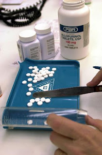
 Pharmacy technician help licensed Pharmacists provide medication and other health care products to patients. Technicians usually perform routine tasks to help prepare prescribed medication, such as counting tablets and labeling bottles. They also perform administrative duties, such as answering phones, stocking shelves, and operating cash registers. Technicians refer any questions regarding prescriptions, drug information, or health matters to a pharmacist.
In hospitals, nursing homes, and assisted-living facilities, technicians have added responsibilities, including reading patients’ charts and preparing the appropriate medication. After the pharmacist checks the prescription for accuracy, the pharmacy technician may deliver it to the patient. The technician then copies the information about the prescribed medication onto the patient’s profile. Technicians also may assemble a 24-hour supply of medicine for every patient. They package and label each dose separately. The packages are then placed in the medicine cabinets of patients until the supervising pharmacist checks them for accuracy, and only then is the medication given to the patients.
Pharmacy aides work closely with pharmacy technicians. They often are clerks or cashiers who primarily answer telephones, handle money, stock shelves, and perform other clerical duties. (See the statement on pharmacy aides elsewhere in the Handbook.) Pharmacy technicians usually perform more complex tasks than pharmacy aides, although in some States their duties and job titles may overlap.
Pharmacy technicians work in clean, organized, well-lighted, and well-ventilated areas. Most of their workday is spent on their feet. They may be required to lift heavy boxes or to use stepladders to retrieve supplies from high shelves.
Technicians work the same hours that Pharmacists work. These may include evenings, nights, weekends, and holidays, particularly in facilities that are open 24 hours a day such as hospitals and some retail pharmacies. As their seniority increases, technicians often acquire increased control over the hours they work. There are many opportunities for part-time work in both retail and hospital settings.
Training
Most pharmacy tech are trained on-the-job, but employers favor applicants who have formal training, certification, or previous experience. Strong customer service skills also are important. Pharmacy technicians may become supervisors, may move into specialty positions or into sales, or may become Pharmacists.
Although most pharmacy technicians receive informal on-the-job training, employers favor those who have completed formal training and certification. However, there are currently few State and no Federal requirements for formal training or certification of pharmacy technicians. Employers who have insufficient resources to give on-the-job training often seek formally educated pharmacy technicians. Formal education programs and certification emphasize the technician’s interest in and dedication to the work. In addition to the military, some hospitals, proprietary schools, vocational or technical colleges, and community colleges offer formal education programs.
Formal pharmacy technician education programs require classroom and laboratory work in a variety of areas, including medical and pharmaceutical terminology, pharmaceutical calculations, pharmacy recordkeeping, pharmaceutical techniques, and pharmacy law and ethics. Technicians also are required to learn medication names, actions, uses, and doses. Many training programs include internships, in which students gain hands-on experience in actual pharmacies. After completion, students receive a diploma, a certificate, or an associate’s degree, depending on the program.
Prospective pharmacy technicians with experience working as an aide in a community pharmacy or volunteering in a hospital may have an advantage. Employers also prefer applicants with experience managing inventories, counting tablets, measuring dosages, and using computers. In addition, a background in chemistry, English, and health education may be beneficial.
Earnings
Median hourly earnings of pharmacy technicians salary in May 2006 were $12.32. The middle 50 percent earned between $10.10 and $14.92. The lowest 10 percent earned less than $8.56, and the highest 10 percent earned more than $17.65. Median hourly earnings in the industries employing the largest numbers of pharmacy technicians in May 2006 were:
| General medical and surgical hospitals | $13.86 |
| Grocery stores | 12.78 |
| Pharmacies and drug stores | 11.50 |
Certified technicians may earn more. Shift differentials for working evenings or weekends also can increase earnings. Some technicians belong to unions representing hospital or grocery store workers.
Interesting ? don't be hesitated. Just visit Medical-Career-Training.com that was designed to provide visitors with the most comprehensive information available on Medical Training programs nationwide, including training for pharmacy technicians . This site provides information on career options for individuals looking to work in the medical field, as well as the degrees available and course offerings at leading institutions. Their school forms are constantly updated to maintain the accuracy and integrity of their partners, and enable you to request information directly from campus and online schools to learn more about their programs.